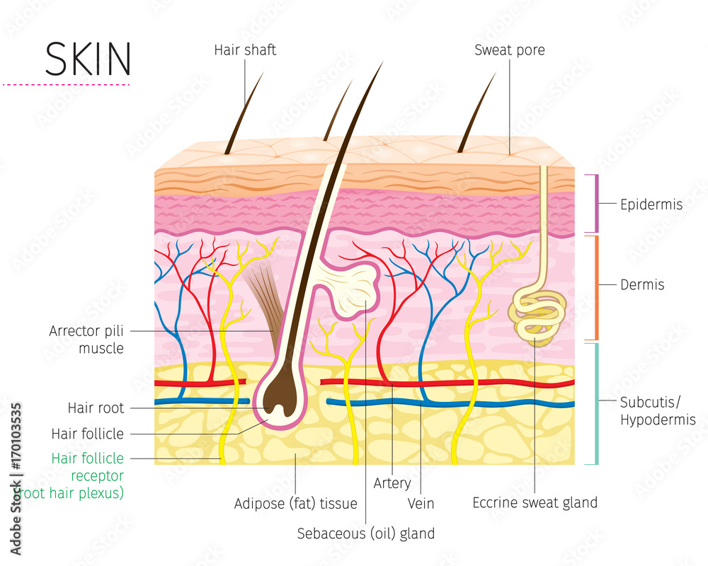
Skin diagram labeled
Skin Diagram The largest organ in the human body is the skin, covering a total area of about 1.8 square meters. The skin is tasked with protecting our body from external elements as well as microbes. Interesting Note:
The Integumentary System (Structure and Function) (Nursing) Part 1
human skin, in human anatomy, the covering, or integument, of the body's surface that both provides protection and receives sensory stimuli from the external environment.The skin consists of three layers of tissue: the epidermis, an outermost layer that contains the primary protective structure, the stratum corneum; the dermis, a fibrous layer that supports and strengthens the epidermis; and.
Skin diagram labeled
Skin anatomy and physiology Hair, skin and nails Wound healing Osmosis High-Yield Notes This Osmosis High-Yield Note provides an overview of Skin Structures essentials. All Osmosis Notes are clearly laid-out and contain striking images, tables, and diagrams to help visual learners understand complex topics quickly and efficiently.
POSTECH UNIVERSITY DEVELOPS 3D BIOPRINTING TECHNIQUE THAT GROWS HUMAN
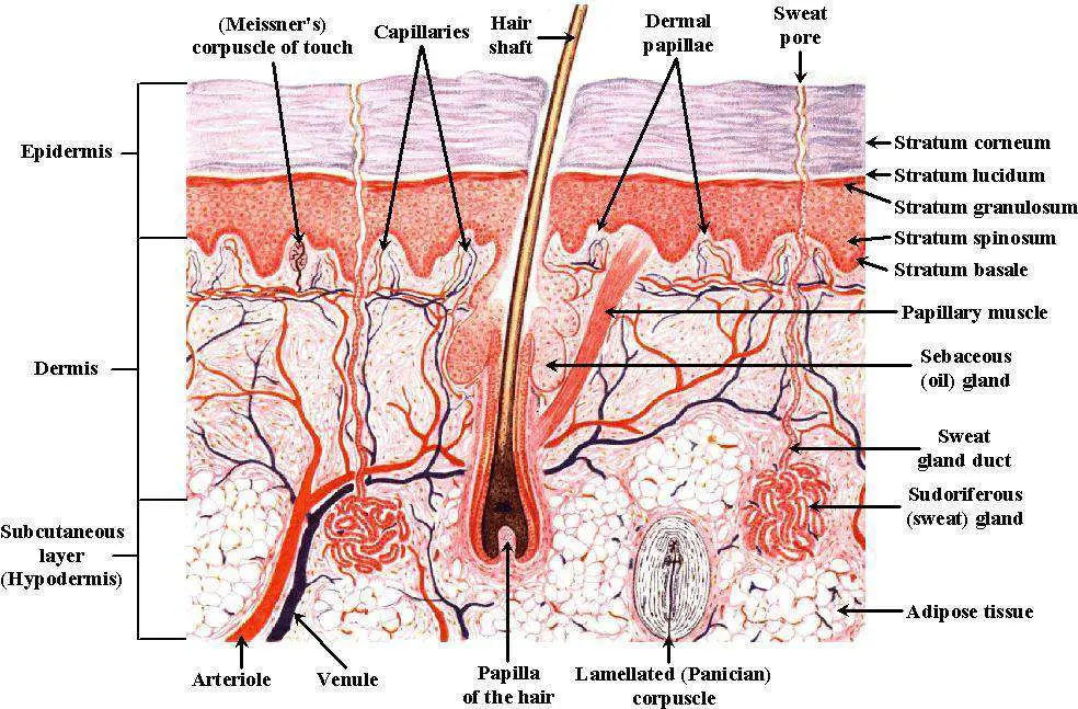
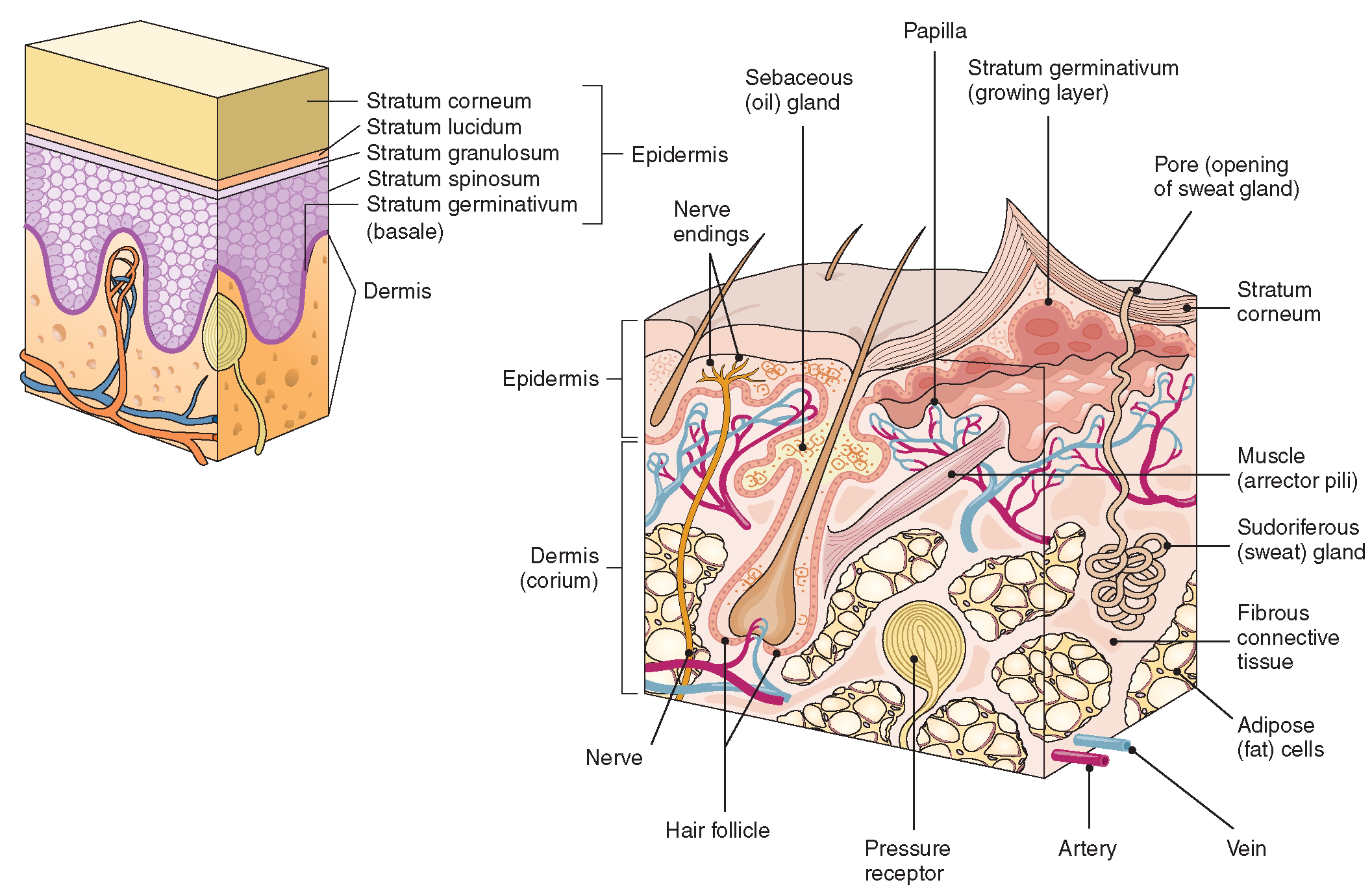
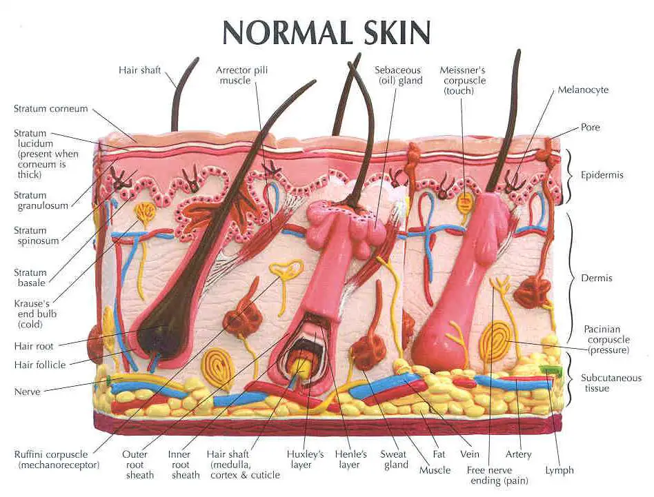
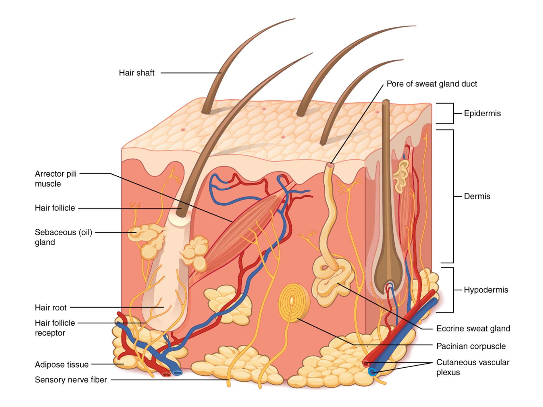
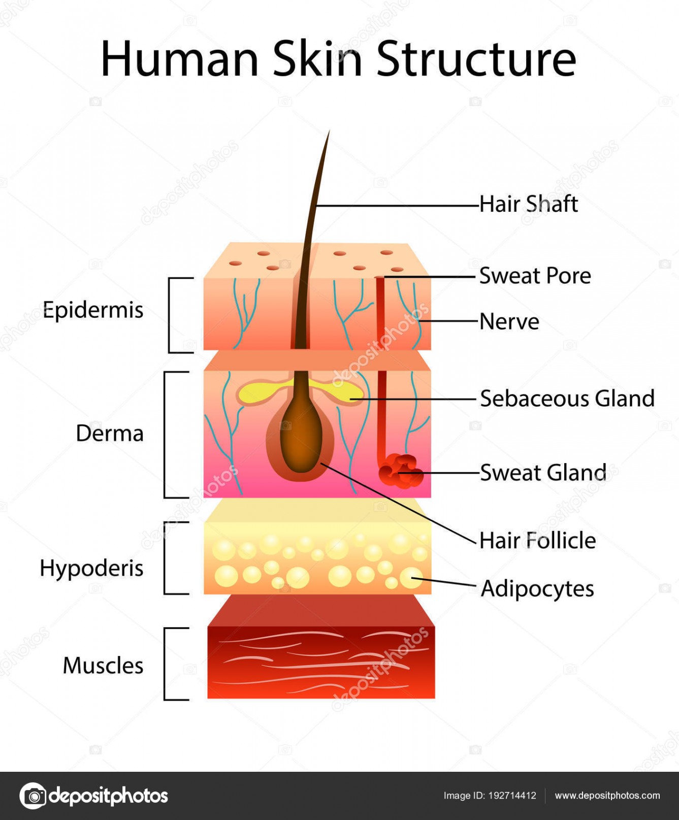
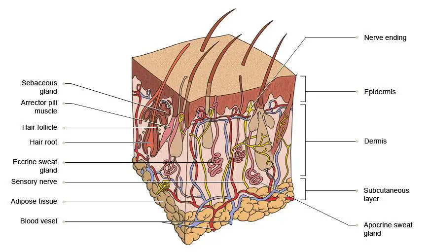
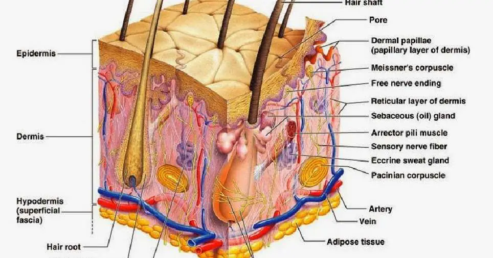
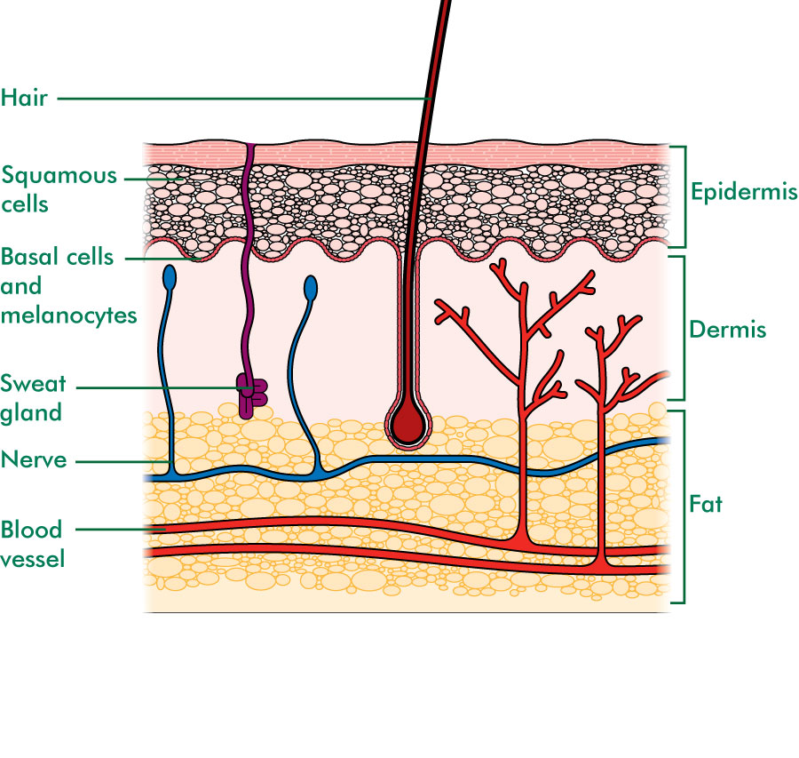
Figure 1. Layers of Skin. The skin is composed of two main layers: the epidermis, made of closely packed epithelial cells, and the dermis, made of dense, irregular connective tissue that houses blood vessels, hair follicles, sweat glands, and other structures.
Skin Diagram Labeled
Epidermis Dermis Subcutaneous fat layer (hypodermis) Each layer has certain functions. Epidermis The epidermis is the thin outer layer of the skin. It consists of 2 primary types of cells: Keratinocytes. Keratinocytes comprise about 90% of the epidermis and are responsible for its structure and barrier functions. Melanocytes.
Labelled Pictures Of Human Skin / skin diagram /medical/anatomy/skin
Dermis. Definition. Fibrous and elastic tissue, provides strength and elasticity to the skin and supports the epidermis, home to hair follicles, glands, nerves etc. Location. Term. Papillary Layer. Definition. Upper dermal layer, provides the epidermis with nutrients and regulates body temperature. Location.

Skin Structure infographic LifeMap Discovery
The skin is the largest organ in the body that covers the entire external surface. It protects the internal organs from germs and thus helps prevent infections. The main functions of the skin include the following: Protecting from water, microorganisms, mechanical and chemical trauma, and damage from UV light.

Anatomy of human skin. The most superficial layer of the skin is the
Skin. As the body's largest organ, skin protects against germs, regulates body temperature and enables touch (tactile) sensations. The skin's main layers include the epidermis, dermis and hypodermis and is prone to many problems, including skin cancer, acne, wrinkles and rashes. Contents Overview Anatomy Conditions and Disorders Care.
Skin diagram labeled
The Epidermis The epidermis is composed of keratinized, stratified squamous epithelium. It is made of four or five layers of epithelial cells, depending on its location in the body. It does not have any blood vessels within it (i.e., it is avascular). Skin that has four layers of cells is referred to as "thin skin."
Skin diagram labeled
The skin is by far the largest organ of the human body, weighing about 10 pounds (4.5 kg) and measuring about 20 square feet (2 square meters) in surface area. It forms the outer covering for the entire body and protects the internal tissues from the external environment. The skin consists of two distinct layers: the epidermis and the dermis.

Cross section anatomy of skin with labels on white background
Key facts about the integumentary system; Skin: Functions: chemical and mechanical barrier, biosynthesis, control of body temperature, sensory Layers: Epidermis (Stratum Basale, Spinosum, Granulosum, Lucidum, Corneum) and dermis (papillary, reticular) Mnemonic: British and Spanish Grannies Love Cornflakes Hair: Types: vellus and terminal Structure: Follicle and bulb (shaft, inner root sheath.

Human Anatomy Diagrams To Label koibana.info Skin anatomy, Human
Dermis Hypodermis Epidermis It is the outermost layer of the skin. The cells in this layer are called keratinocytes. The keratinocytes are composed of a protein called keratin. Keratin strengthens the skin and makes it waterproof. Melanocytes that produce melanin are also present in this layer.
Skin diagram to label Labelled diagram
From top, LM × 40, LM × 40. (Micrographs provided by the Regents of University of Michigan Medical School © 2012) The cells in all of the layers except the stratum basale are called keratinocytes. A keratinocyte is a cell that manufactures and stores the protein keratin.
The skin Understanding cancer Macmillan Cancer Support
The skin is the body's largest and primary protective organ, covering its entire external surface and serving as a first-order physical barrier against the environment. Its functions include temperature regulation and protection against ultraviolet (UV) light, trauma, pathogens, microorganisms, and toxins. The skin also plays a role in immunologic surveillance, sensory perception, control of.

human skin cells labeled Google Search Subcutaneous tissue, Skin
The Epidermis The Dermis Hypodermis The number of skin layers that exists depends on how you count them. You have three main layers of skin—the epidermis , dermis, and hypodermis (subcutaneous tissue). Within these layers are additional layers. If you count the layers within the layers, the skin has eight or even 10 layers.

Skin Diagram Labeled
Introduction Skin is the largest organ in the body and covers the body's entire external surface. It is made up of three layers, the epidermis, dermis, and the hypodermis, all three of which vary significantly in their anatomy and function.